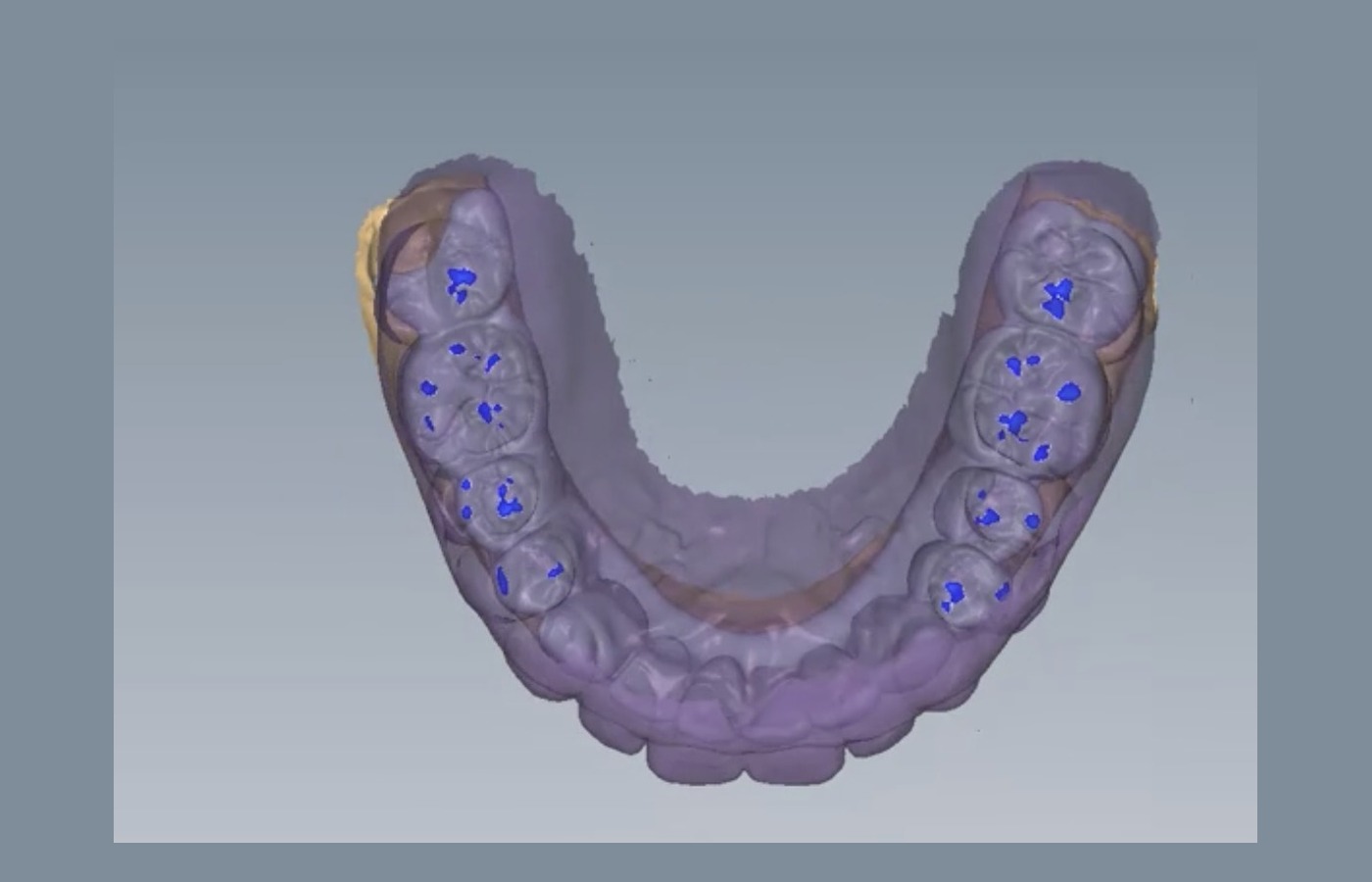
Joint-guided mandibular position planning for clinician-selected TMJ-sensitive restorative, orthodontic and full-mouth rehabilitation cases.
AxisGuide is a staged digital planning workflow that uses MRI measurement of disc displacement amount and direction, CBCT and intraoral scans to simulate a proposed mandibular position. A 3D-printed localizator is designed from the interarch space at that position, so the clinician can review joint response, disc-condyle relationship and symptom response before provisional occlusal reconstruction is trialed.
Condylar vector simulationSkull and dentition simulation
Splints & Deprogrammers
Appliance CAD
For straightforward splint or functional appliance requests where the bite position is already defined by the clinician.
Typical output
Michigan splints, deprogrammers, stabilization splints, night guards, bite ramps, and production files.
AxisGuide
Joint-position-first planning
For complex cases where the mandibular position itself must be evaluated before orthodontic or restorative reconstruction.
Typical output
MRI displacement-vector review, mandibular movement simulation, 3D-printed localizator design, verification references, provisional occlusal setup, and orthodontic or restorative reconstruction references.
Technique logic
MRI-guided thinking, translated into a collaborative digital workflow.
The workflow does not start from a conventional bite rim. MRI measurements define the disc displacement vector, mandibular movement is simulated digitally, and a printed localizator carries that proposed position for clinician-led symptom and MRI verification before a provisional occlusal reconstruction is trialed.
01
Record integration
MRI, CBCT, intraoral scans, clinical symptoms, photos and rehabilitation goals are aligned into one planning context.
02
MRI vector measurement
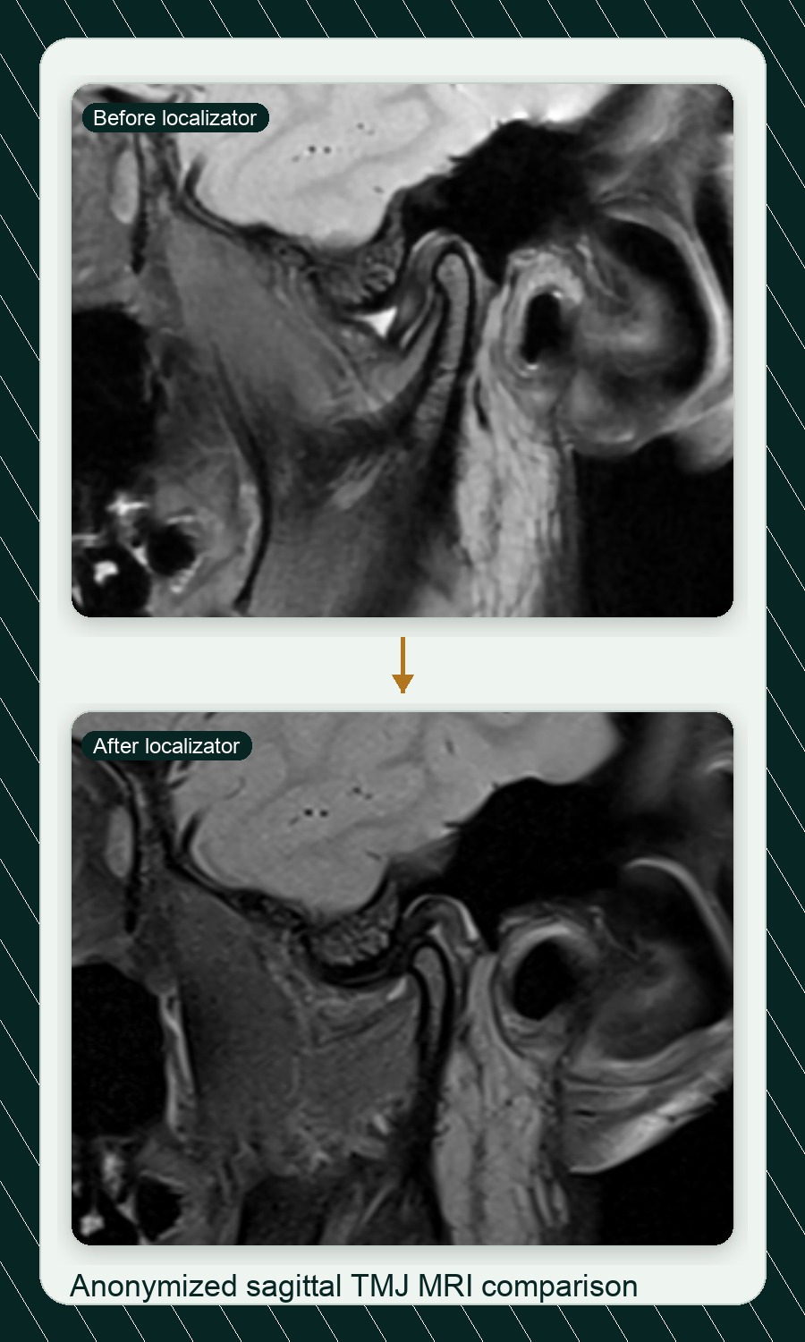
The amount and direction of disc displacement are measured from MRI and translated into a planning vector for the proposed mandibular position.
03
Mandibular movement simulation
The mandible is simulated into the proposed position, with interarch space, condylar references and occlusal clearance reviewed digitally.
04
Localizator verification
A 3D-printed localizator is designed from that interarch space so the clinician can check disc-condyle relationship, MRI verification and symptoms.
05
Provisional occlusal trial
If verification is stable, provisional teeth or a temporary occlusal reconstruction can test MIP contacts, dynamic occlusion, canine guidance and anterior guidance.
06
Orthodontic or restorative reconstruction
The verified position can then guide orthodontic occlusal reconstruction, definitive restorative reconstruction, or a combined treatment sequence.
Localizator CAD detail
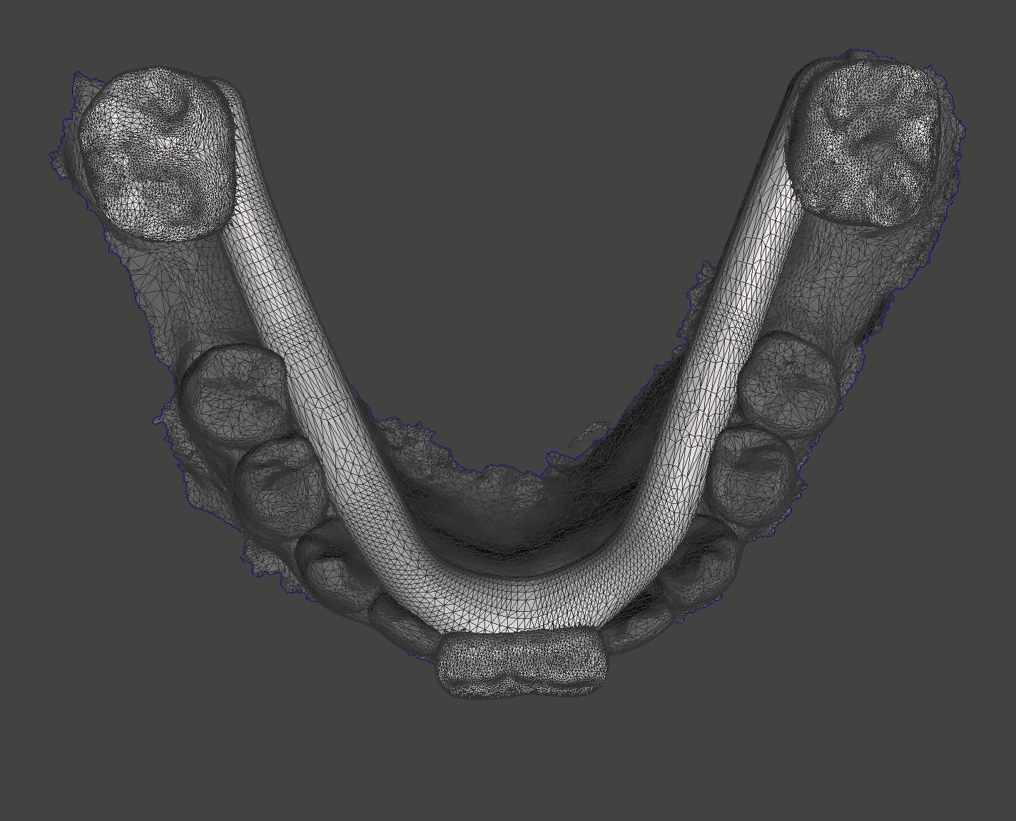
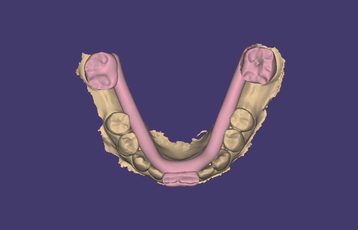
The planned interarch space is translated into a CAD-designed position carrier for clinical verification, not a generic splint.
Digital position record
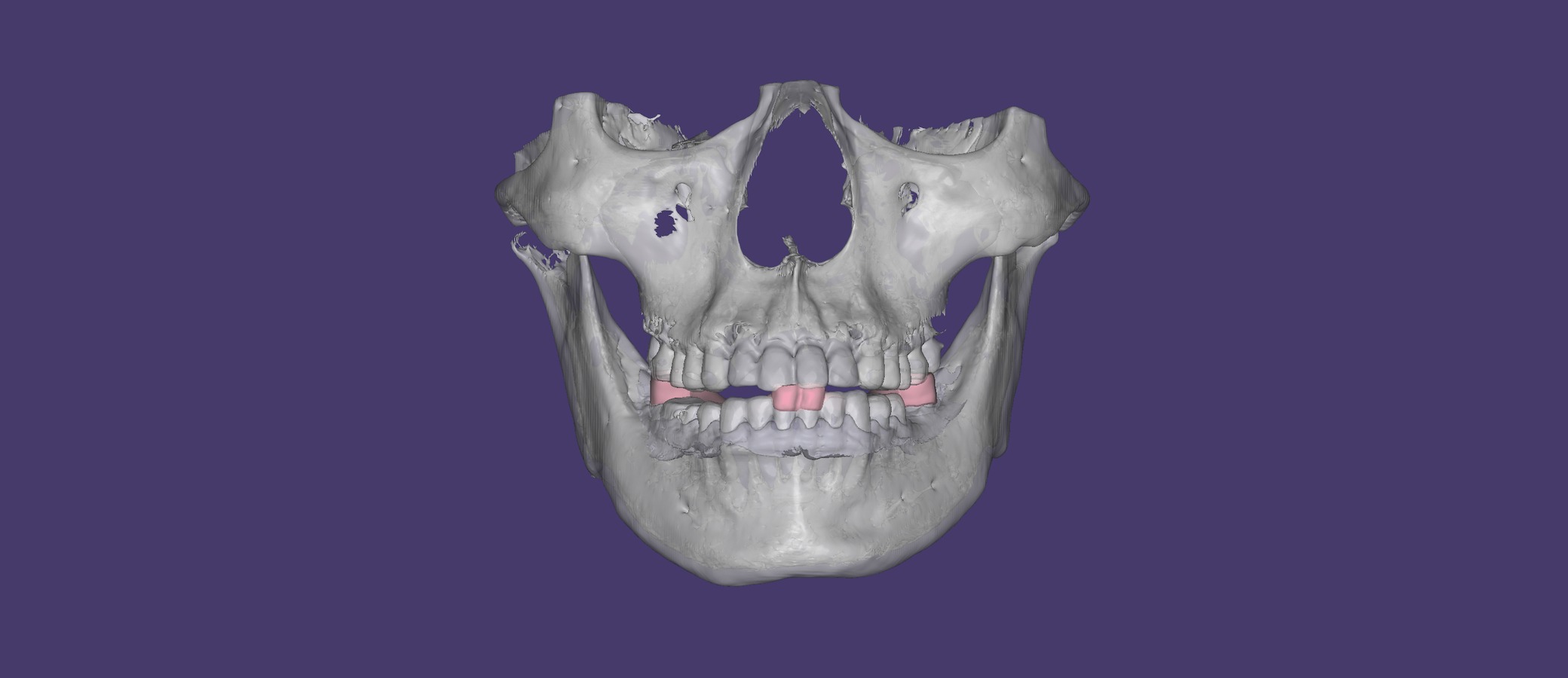
The proposed mandibular position is documented against skull references, intraoral scans and condylar references before the localizator is fabricated.
Provisional occlusal trial
The provisional setup can be reviewed against planned MIP static contacts before dynamic occlusion, canine guidance and anterior guidance are refined.
Best-fit cases
Full-mouth rehabilitation where the starting bite is unreliable
Orthodontic-restorative cases requiring mandibular position coordination
Worn dentition, VDO change, deep bite, crossbite or unstable occlusal records
Clinician-selected TMJ cases with limited opening, joint pain, joint sounds or deviation on opening
Facial asymmetry, mandibular deviation or mandibular retrusion requiring interdisciplinary planning
TMJ-sensitive cases where clinicians want imaging-informed planning support
Planning deliverables
Record checklist and case-readiness review
MRI displacement-vector planning references
Digital mandibular movement simulation
3D-printed localizator CAD support
Provisional occlusal reconstruction references
Orthodontic or restorative reconstruction sequencing support
Clinical boundary
Diagnosis and treatment decisions remain with the clinician
Planning and verification are reviewed case by case with the treating team
Disc-condyle relationship, symptoms and imaging findings are verified clinically when indicated
Degenerated joints or non-reducible disc conditions require careful case selection
Definitive orthodontic or restorative sequencing remains clinician-led
Positioning noteAxisGuide is an advanced digital planning and collaboration service. It supports professional review and treatment planning while diagnosis, therapy selection and outcome evaluation remain under clinician control.
Discuss an AxisGuide caseShare anonymized TMJ MRI, CBCT/DICOM, intraoral scans, current mandibular position records and the rehabilitation goal so the scope can be reviewed before quote.